
Hexarelin Acetate Research Peptide: Separating GHS-R1a From CD36
Tetrava Labs Editorial Team10 min read
Hexarelin acetate engages GHS-R1a and binds CD36, but those targets do not explain the same endpoints. This research guide separates endocrine assays, cardiac models, salt mass, and analytical controls.

Introduction
The hexarelin acetate research peptide is a synthetic growth hormone secretagogue with an awkward experimental property: the same molecule can engage the growth hormone secretagogue receptor type 1a (GHS-R1a) and bind the scavenger receptor CD36. Those targets occupy different signaling contexts. A change in growth hormone release, coronary perfusion pressure, oxidized lipoprotein uptake, or cardiomyocyte survival cannot be assigned to one mechanism simply because hexarelin was present.
This dual-target pharmacology makes clean controls more important than a long endpoint panel. Researchers must separate receptor occupancy from downstream phenotype, distinguish peptide base from acetate salt, and control for the nonstandard residues that give hexarelin its sequence-level behavior. Hexarelin is sold for research use only. It is not approved for human consumption, diagnosis, therapy, or veterinary use.
What is hexarelin acetate?
Hexarelin, also called examorelin in parts of the literature, is a six-residue synthetic peptide in the growth hormone-releasing peptide family. Its sequence is commonly written His-D-2-methyl-Trp-Ala-Trp-D-Phe-Lys-NH2. The D-configured residues, methylated tryptophan, and amidated C-terminus are deliberate chemical features rather than ordinary protein translation products.
Those nonstandard features affect more than a sequence diagram. They can change protease susceptibility, chromatographic retention, ionization behavior, receptor affinity, and the fragmentation pattern expected during mass spectrometry. An identity method should therefore confirm the intact mass and, where feasible, sequence-informative fragments. A generic amino-acid assay or retention-time match alone is weak evidence of identity.
What acetate means on the label
"Acetate" identifies a counterion paired with protonated sites on the peptide. It does not mean that an acetyl group has been covalently attached to the sequence. Hexarelin base mass and hexarelin acetate mass are therefore different accounting quantities: the salt includes acetate counterion mass, while the peptide base does not.
Counterion stoichiometry may vary with purification and isolation conditions. Residual water and any declared excipients can add further mass to a lyophilized preparation. A study that normalizes concentration by gross vial mass without a peptide-content assay may compare unequal molar amounts. Report whether calculations use as-is material, anhydrous salt, or peptide-equivalent content, then preserve that convention across groups.

How does hexarelin work through GHS-R1a?
GHS-R1a is a G-protein-coupled receptor expressed in pituitary, hypothalamic, and peripheral tissues. Agonist binding can recruit phospholipase C-linked signaling, increase intracellular calcium, and promote secretory responses in suitable cellular systems. In endocrine experiments, growth hormone output is the familiar endpoint, but it is not a direct receptor-binding measurement. Somatostatin tone, growth hormone-releasing hormone input, receptor reserve, and feedback can all change the observed response.
The endocrine profile is broader than growth hormone alone. A human endocrine study reported activation of the hypothalamo-pituitary-adrenal axis and used hormone-pattern analysis to support a role for arginine vasopressin. That finding is useful as a warning about assay interpretation: a secretagogue can alter several endocrine outputs through network effects. It does not establish that every downstream signal comes from direct action in the measured tissue.
Potency is not the same as sustained response
A low EC50 in one signaling assay does not predict a durable endocrine phenotype. GHS-R1a can display constitutive activity, ligand-dependent internalization, pathway bias, and time-dependent desensitization. Compare full concentration-response curves, maximal efficacy, onset, washout, and repeat-challenge behavior. A single peak value can make a high-efficacy ligand look preferable while concealing faster signal decay.
GHS-R1a vs CD36: two targets, different claims
CD36 is a multifunctional scavenger receptor found in cardiomyocytes, microvascular endothelium, macrophages, and other cell types. In a 2002 cardiac membrane study, investigators identified an approximately 84 kDa hexarelin-binding protein as CD36. Hexarelin increased coronary perfusion pressure in isolated hearts, and that response was absent in two CD36-deficient models. Within that specific preparation and endpoint, CD36 loss supplied strong mechanistic evidence.
A later photoaffinity cross-linking study mapped a hexarelin-binding region to CD36 residues Asn132-Glu177, overlapping the reported oxidized LDL-binding region. The authors proposed that competition at this site might affect modified lipoprotein uptake. Binding-site overlap supports a testable mechanism. It does not, by itself, prove a cardiovascular outcome or specify what happens in a whole organism.
Cardiac results should not be labeled CD36-only. In a 2017 rat ischemia-reperfusion study, hexarelin was associated with altered IL-1beta and IL-1 receptor antagonist expression, lower oxidative-stress readouts, and improved cardiac measures. A GHS-R antagonist neutralized part of the signaling result, supporting GHS-R1a involvement in that model. The study discussed both GHS-R1a and CD36, yet its antagonist evidence prevents a clean CD36-only interpretation.

Hexarelin vs ipamorelin: what a fair comparison requires
Hexarelin and ipamorelin are often reduced to a ranking of growth hormone release. That is an incomplete comparison. As the ipamorelin comparison guide explains, ligands can differ in receptor family, selectivity, and endpoint behavior. The compounds should be evaluated at matched molar concentrations and under the same receptor-expression conditions before potency claims are compared.
Four dimensions belong in the comparison: GHS-R1a potency, maximal efficacy, off-target selectivity, and desensitization across time. Endpoint controls matter just as much. Receptor-proximal calcium flux, beta-arrestin recruitment, hormone release, and tissue phenotype answer different questions. If hexarelin is tested in a CD36-positive cell while ipamorelin is tested only in a recombinant GHS-R1a system, the result confounds ligand identity with model biology.
Study design for dual-target pharmacology
A defensible design uses orthogonal perturbations. One inhibitor is rarely enough because receptor antagonists can have incomplete occupancy or off-target effects. Pair pharmacology with genetic loss of function, then restore the receptor when the model permits. The following controls separate target engagement from downstream association:
- Use GHSR knockout or knockdown cells beside matched wild-type cells, with a validated GHS-R1a antagonist and a receptor-rescue condition.
- Use CD36-null or silenced models, a CD36-blocking reagent where validated, and rescue with controlled CD36 expression.
- Include ghrelin or another characterized GHS-R1a agonist as a pathway comparator, plus a CD36-binding comparator that lacks the same endocrine profile.
- Measure receptor-proximal events before distal outcomes. Confirm target engagement, then record calcium, second messengers, transcriptional changes, and the tissue endpoint on a defined timeline.
- Predefine whether the experiment tests endocrine secretion, lipid handling, vascular tone, inflammatory signaling, or cell survival. Avoid treating a panel of loosely related changes as one mechanism.
Time and matrix controls deserve equal attention. Peptide adsorption to plastic, degradation in serum-containing media, and freeze-thaw history can shift free concentration. Analyze stock stability under the actual assay conditions. A nominal concentration is not proof that intact hexarelin reached either receptor.
Build an endpoint matrix before collecting data
Map each readout to the evidence needed for attribution. A GHS-R1a binding or reporter assay establishes receptor-proximal activity, but it says little about CD36. Photoaffinity labeling, competition binding, or a validated CD36-dependent uptake assay addresses the second target. Hormone secretion, cytokine expression, coronary pressure, and cell survival sit farther downstream and need a chain of controls between receptor engagement and phenotype.
Use a two-by-two target design when both receptors are present: GHS-R1a intact or disrupted, crossed with CD36 intact or disrupted. This reveals whether either receptor is sufficient, whether both are required, or whether one receptor changes the apparent contribution of the other. Add vehicle, untreated, and positive-control groups to detect solvent effects and assay drift. Randomize sample processing order and blind image or pathology scoring where those endpoints are used.
Concentration selection should come from a verified range-finding experiment, not from an unrelated tissue paper. Confirm viability and nonspecific membrane effects at the upper range. Record biological and technical replicate counts separately. If several endpoints are tested, define the primary endpoint and multiplicity plan before viewing the results; otherwise, a noisy secondary signal can become the story after the fact.
What the COA should establish
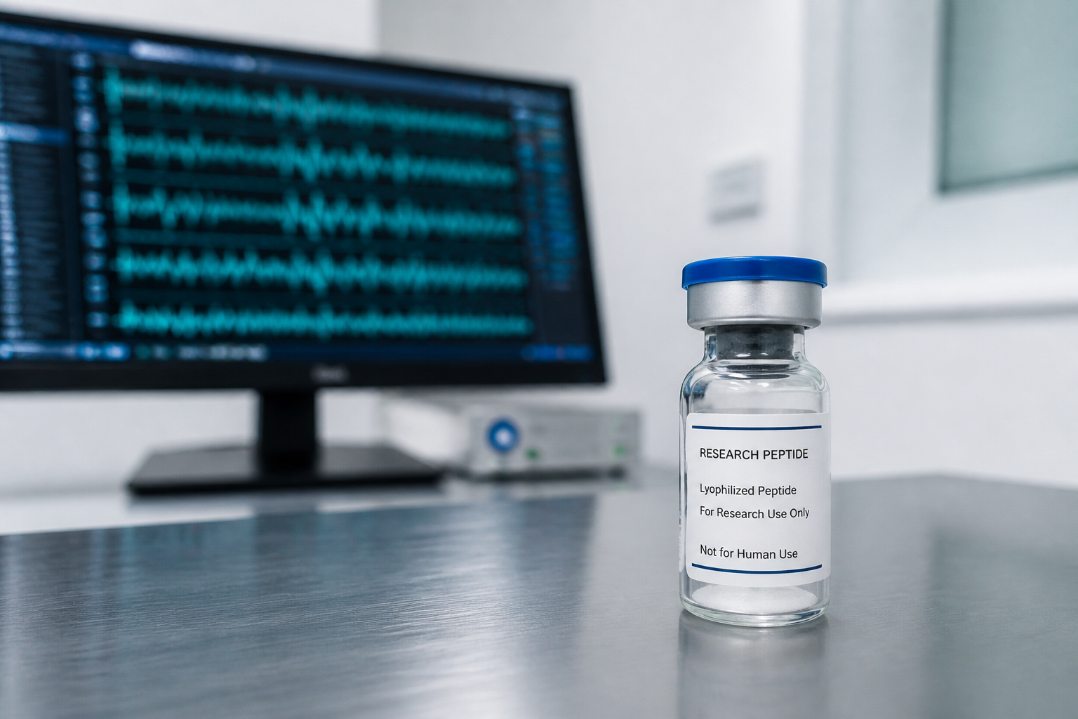
A lot-linked COA should connect the tested sample to the vial used in the experiment. At minimum, review chromatographic purity, mass-spectrometric identity, lot number, test date, and the laboratory responsible for each method. Purity area percent is not peptide content, and neither measurement establishes sterility or endotoxin status unless those tests are reported separately. Lot records published by Tetrava Labs should match the vial label before any assay begins.
For acetate material, seek counterion data and water content when quantitative molarity matters. Review the chromatogram rather than accepting a rounded purity claim. Unresolved shoulders, integration choices, and a method with no stated wavelength or column conditions limit reproducibility. For cell work, endotoxin can mimic inflammatory biology; a clean HPLC trace does not rule it out.
Interpretation boundaries
Three common shortcuts fail. First, growth hormone release does not prove direct action in every tissue. Second, CD36 binding does not make every cardiovascular observation CD36-mediated. Third, a rat ischemia-reperfusion result does not establish a human therapeutic effect. Each inference needs its own receptor control, species context, and endpoint validation.
The useful question is narrower: which target is required for a defined response under stated conditions? A result that survives GHS-R1a loss but disappears with CD36 loss supports one path. A result blocked by a GHS-R1a antagonist and preserved after CD36 loss supports another. Mixed attenuation may be biologically real, but it demands a factorial design rather than a single-target story.
Two receptors, one peptide, no shortcuts. Every hexarelin result has to say which target did the work.
Conclusion
Hexarelin acetate is experimentally interesting because its pharmacology resists a one-receptor explanation. GHS-R1a provides a defined route into endocrine and some cardiac signaling, while CD36 binding supports separate hypotheses in vascular and lipid-handling models. Salt accounting, sequence-aware identity testing, receptor knockout or antagonist controls, and matched time-course endpoints turn that ambiguity into a workable study. Keep every inference tied to the model that produced it, and keep the material restricted to laboratory research.
Frequently Asked Questions (FAQ)
What is hexarelin acetate? A six-residue synthetic growth hormone secretagogue peptide, also called examorelin, supplied as an acetate salt for laboratory research use only.
How does hexarelin work? Primarily through the growth hormone secretagogue receptor GHS-R1a in endocrine tissue, though it also binds the scavenger receptor CD36 in cardiac and vascular tissue through a separate pathway.
What is the difference between the GHS-R1a and CD36 effects? GHS-R1a activation is linked to growth hormone secretion. CD36 binding is linked to coronary perfusion and lipid-handling effects in cardiac tissue. A result needs a receptor-specific control before either target can be credited.
How does hexarelin compare to ipamorelin? Both are GHS-R1a agonists, but they differ in selectivity and off-target binding. Hexarelin's added CD36 activity means a fair comparison has to control for which receptor produced the observed effect.
References
- Korbonits M et al. (1999). The growth hormone secretagogue hexarelin stimulates the hypothalamo-pituitary-adrenal axis via arginine vasopressin. The Journal of Clinical Endocrinology & Metabolism
- Bodart V et al. (2002). CD36 mediates the cardiovascular action of growth hormone-releasing peptides in the heart. Circulation Research
- Demers A et al. (2004). Identification of the growth hormone-releasing peptide binding site in CD36: a photoaffinity cross-linking study. The Biochemical Journal
- Xu X et al. (2017). The growth hormone secretagogue hexarelin protects rat cardiomyocytes from in vivo ischemia/reperfusion injury through interleukin-1 signaling pathway. International Heart Journal

Editorial Team, Tetrava Labs
Content published by Tetrava Labs is compiled and fact-checked using peer-reviewed scientific literature, HPLC-MS Certificates of Analysis (COA), and primary biochemical data. Research use only.
Research Use Only Disclaimer
All products are intended for laboratory research purposes only. Not approved for human consumption, diagnostic use, or therapeutic applications. By purchasing, you confirm you are a qualified research professional.
Related articles
Selank Research Peptide: Sequence, Mechanisms, and Evidence
A source-led review of Selank's TKPRPGP sequence, tuftsin lineage, GABA and BDNF findings, evidence limits, assay design, and COA requirements.

TUDCA Research Compound: Chemistry, ER Stress, and the Chemical Chaperone Question
TUDCA is a taurine-conjugated bile acid, not a peptide. This research review separates UPR marker changes from direct protein-folding evidence and examines a 2025 challenge to the chemical chaperone label.
Follistatin 315 Research Peptide: Protein Identity, Isoforms, and COA Verification
Follistatin 315 is a 315-amino-acid secreted glycoprotein, not an undefined short peptide fragment. Learn how FST315 differs from FST288 and FST344, how follistatin sequesters activin and myostatin, and what analytical evidence a defensible COA must provide.